Research Overview
The development of medical optical imaging techniques has provided effective research tools to investigate many fundamental processes in the life sciences. In most cases, optical detection requires exogenous agent to either provide a signal readout or enhance image contrast. Our group develops a variety of imaging probes and multifunctional platforms to explore pressing biomedical problems. Our current research is focused on the development of fluorescent probes, biosensors, and spectroscopic and imaging techniques for biomedical applications.
Development of bright semiconducting polymer dots for subcellular labeling and super-resolution imaging.
Demonstration of robust polymer dot sensors for in vitro assays and in vivo dynamic glucose monitoring.
Development of photosensitizing polymer dots and enzyme-enhanced phototherapy for cancer treatment.
Design and Construction of spectroscopic and imaging instrumentation.
Semiconducting polymer dots for biomedical applications.
Research Project
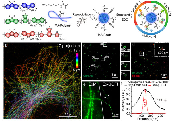
Development of bright semiconducting polymer dots for subcellular labeling and super-resolution imaging
We developed two types of small photoblinking Pdots with high brightness, strong photostability, and favorable biocompatibility. Furthermore, we have demonstrated their excellent performance as fluorescent probes for super-resolution optical fluctuation imaging (SOFI) nanoscopy, a typical blinking-based statistical super-resolution imaging technique. Benefiting from the 3D PSF modulation (enhanced resolution in three-dimensions) and background reducing capacity of SOFI, we achieved background-free SOFI super-resolution imaging of several subcellular structures labeled with small photoblinking Pdots in both two and three dimensions.
Research Project
Development of photosensitizing polymer dots and enzyme-enhanced phototherapy for cancer treatment
We constructed a nanoparticle platform by covalent conjugation of glucose oxidase (GOx) to small polymer dots, which could be persistently immobilized into a tumor. While the malignant tumors have high glucose uptake, the GOx efficiently catalyzes the glucose oxidation with simultaneous generation of H2O2. Under light irradiation, the in situ generated H2O2 was photolyzed to produce hydroxyl radical, the most reactive oxygen species, for killing cancer cells. The remarkable inhibition of tumor growth was observed in xenograft-bearing mice, indicating the promise of the EEPT approach for cancer therapeutics.
Research Project
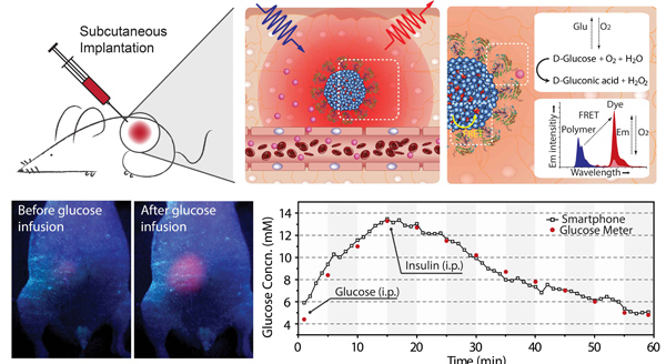
Demonstration of robust polymer dot sensors for in vitro assays and in vivo dynamic glucose monitoring
We describe in vivo real-time dynamic monitoring of small molecules by a luminescent polymer-dot oxygen transducer. The optical transducer combined with an oxygen-consuming enzyme can sensitively detect small-molecule substrates as the enzyme-catalyzed reaction depletes its internal oxygen reservoir in the presence of small molecules. We exemplify this detection strategy by using glucose-oxidase-functionalized polymer dots, yielding high selectivity, large dynamic range, and reversible glucose detection in cell and tissue environments. We further developed an image processing algorithm and a software application that was installed on a smartphone. Wireless and real-time dynamic glucose monitoring in live mice was demonstrated with the smartphone and the implanted transducer.